Neurochemical Alterations in Bilateral DLPFC after Structure Learning Training in Healthy Adults
Abstract
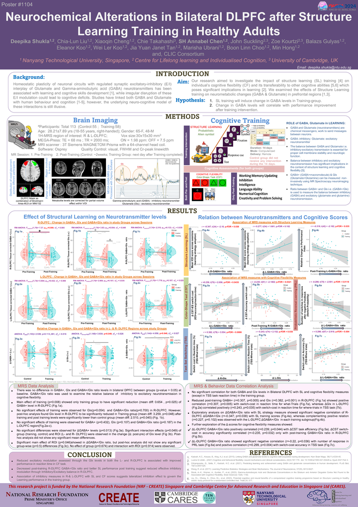
Introduction:
Structural learning integrates ‘Learning to learn’ approach of individual’s abilities to extract underlying pattern and develop rules to adapt new changes through cognitive flexibility. Homeostatic plasticity in neuronal circuits is crucial for critical learning and depends upon coordinated modulation of synaptic excitation and inhibition through Glutamate and GABA interactions. Disruption in this coordinated neurotransmitter’s interplay triggers cognitive deficits, while adaptive modulation contributes to relearning capacity. Studies reported associative interaction with learning and cognitive skills development with neurotransmitters, but the underlying neuro-cognitive model of these interactions is illusive. Using controlled SL training intervention, we aim to investigate the effect of learning in both the neuronal and behavioral levels to assess its transferability to other cognitive abilities.
Methods:
113 healthy volunteers aged between 18-55 years were pseudo-randomized to control and training groups matching with age, gender and intelligence. T-group underwent 2-week SL training. Out of the 113, 106 participants completed with post-session magnetic resonance imaging, of which 7 withdrew or dropped out of the study. All MR scans were performed in 3T Siemens MAGNETOM Prisma MRI scanner with a 64-channel head coil. All participants consented to Cognitive testing and MRI sessions with ethics approval from NTU-IRB. MR spectroscopy for GABA quantitation in bilateral left- and right-dorsolateral prefrontal cortex were performed at two different time points of pre- and post- SL training sessions along with cognitiveassessments. Each MR session included 3D T1-MPRAGE and 1H-MEGA-PRESS MRS with one unsuppressed water spectra of Navg is 4. Voxels were placed close to middle frontal gyrus maximizing gray matter. Manual shimming resulted linewidth less than 16 Hz. MRS data in BIDS structure was applied for pre-processing and Osprey was used for quantitation of GABA (GABA and macromolecule) and Glx (Glutamate and glutamine). Quality check for MRS data included visual artefacts, head movements, broad Creatine (Cr) linewidth in the OFF-spectra, and poor fitting.
Results:
Tissue corrected GABA and Glx levels in both left and right DLPFC of study groups did not differ at pre-training stage. After training, the T-group showed significant reduction in R-DLPFC Glx compared to C-group. Paired comparison between sessions showed significant decrease in post-training R-DLPFC GABA in T-group but not in C-group. No significant difference was observed for L-DLPFC GABA, Glx and GABA-Glx ratio across groups and sessions. MRS measures did not relate to SL test-scores. However, R-DLPFC Glx in the T-group correlated positively with switch-cost reaction time between shift-repeat trials of color-shape task, indicating reduced Glx levels in the R-DLPFC relates to short reaction time in the T-group. GABA-Glx ratio in T-group showed significant positive relation with probability shift measure levels in contrast to negative relation observed in C-group. A strategy shifting ability in the T-group is observed in CF, and other cognitive domains (i.e. working memory, inhibition, and non-verbal intelligence) in contrast to C-group.
Deepika Shukla
Research Fellow
Dr Deepika Shukla is a Research Fellow in the Centre for Lifelong Learning and Individualised Cognition (CLIC) Neuroimaging team.
Annabel Chen
Professor of Psychology
Lab Director
Dr. SH Annabel Chen is a clinical neuropsychologist, and currently a Faculty member of Psychology at the School of Social Sciences.
Koo Wei Ler
Alumni
Koo Wei Ler is a research assistant in the Centre for Lifelong Learning and Individualised Cognition (CLIC).